Answer DV Case 6
| This page is part of De Voogt Archive - Cases
Previous Case: DVA Case 5 | Next Case: DVA Case 7 |
Questions
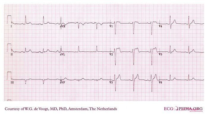
This is a tracing of a 52 year old male, with viral infection and high fever (40° C), who was admitted to the hospital with syncopal attacks, loss of consciousness and seizures.
One of these attacks was recorded and the ECG monitor tracing showed Ventricular Fibrillation (Torsades de pointes).
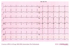
In the VF protocol, amiodarone iv was infused. This produced the ECG on the right, but VF was repeatedly seen.
- What is your diagnoses?
- What is your management?
Answer
The original ECG, is the typical cove type Brugada syndrome ECG. Often this type of ECG is confusing when an acute anterior myocardial infarct is suspected. This patient received amiodarone. The prolongation of the QT interval is noticeable on the second tracing. Also note the rapid change in QRS configuration. The configuration however, is still suggestive for Brugada syndrome. Proper treatment of the repetitive VT induced by fever, was instituted by sedation and cooling of the patient. Amiodarone was stopped as it is not the drug of choice in Brugada syndrome. When temperature was lowered to < 37° C , VF did not recur. A week later an ICD was implanted.

