Answer DV Case 4: Difference between revisions
Jump to navigation
Jump to search
m (New page: # The rhythm shows inverted P-waves in the inferior leads indicative of atrial rhythm. # The answer on this question is difficult. An atrial rhythm can be the result of SA block. This ECG ...) |
mNo edit summary |
||
| Line 1: | Line 1: | ||
{{Case| | |||
|previouspage= / | |||
|previousname= / | |||
|nextpage= / | |||
|nextname= / | |||
}} | |||
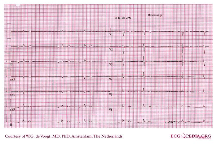
[[Image:DVA0004.jpg|700px|thumb|left|DV Case 4. Click on image for enlargement.]] | |||
{{clr}} | |||
==Questions== | |||
This ECG shows pauses in the heart rhythm. The patient felt light headiness | |||
#What is the base rhythm? | |||
#Is there evidence for sino atrial block? | |||
#What is the cause of the pauses in the heart rhythm? | |||
#Is there a pacemaker indication? | |||
#what is your next step in your workup? | |||
==Answers== | |||
# The rhythm shows inverted P-waves in the inferior leads indicative of atrial rhythm. | # The rhythm shows inverted P-waves in the inferior leads indicative of atrial rhythm. | ||
# The answer on this question is difficult. An atrial rhythm can be the result of SA block. This ECG however cannot produce the proper answer. The use of beta-blockers for instance, could cause a marked lowering of the sinus rhythm, allowing for an atrial escape rhythm, which is slow as well. | # The answer on this question is difficult. An atrial rhythm can be the result of SA block. This ECG however cannot produce the proper answer. The use of beta-blockers for instance, could cause a marked lowering of the sinus rhythm, allowing for an atrial escape rhythm, which is slow as well. | ||
Revision as of 21:50, 10 November 2008
| This page is part of Cases and Examples |
Questions
This ECG shows pauses in the heart rhythm. The patient felt light headiness
- What is the base rhythm?
- Is there evidence for sino atrial block?
- What is the cause of the pauses in the heart rhythm?
- Is there a pacemaker indication?
- what is your next step in your workup?
Answers
- The rhythm shows inverted P-waves in the inferior leads indicative of atrial rhythm.
- The answer on this question is difficult. An atrial rhythm can be the result of SA block. This ECG however cannot produce the proper answer. The use of beta-blockers for instance, could cause a marked lowering of the sinus rhythm, allowing for an atrial escape rhythm, which is slow as well.
- The cause of the pauses in the atrial rhythm are atrial premature beats, blocking the exit of the atrial prevailing atrial rhythms. These atrial premature beats can be appreciated when looking at the T-waves of the conducted beats preceding the pause.
- This question cannot be answered on this ECG. Pacing could be indicated as the patient has dizzy spells. However, blocked atrial beats per se are no pacemaker indication.
- The proper workup could be: Stopping medication that lowers the sinus node frequency (B-blockers, funny current blockers, calcium channel blockers).
And in any case a long term ECG recording. Measuring the sinus node recovery time is questionable.
