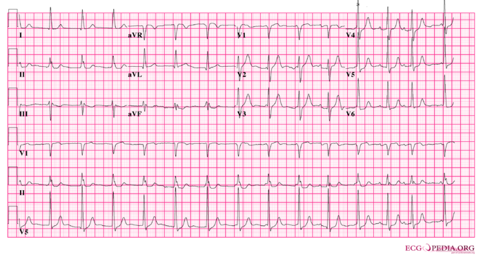
Answer Case 5
Jump to navigation
Jump to search
| This page is part of Cases and Examples |
Try to interprete this ECG using the 7+2 step method
Answer
- Following the 7+2 steps:
- Rhythm
- The ECG shows a regular rhythm, but the last couple of beats are faster. Between these last beats clear P waves are discernable. De distance between the P waves and QRS complexes changes and there seems to be no relation between the two: AV dissociation. As there are no P waves preceding the first QRS complexes the rhythm must be of nodal origin in competition with sinusrhythm. The 10th, 12th and 13th beats are preced by a P wave, here the sinusrhythm has taken over from the nodal rhythm.
- Heart rate
- around 80 bpm
- Conduction (PQ,QRS,QT)
- PQ: not applicable. QRS: 110ms QT: 380ms
- Heartaxis
- Positive in I and II, negative in III. Slightly positive in AVF. An intermediate heart axis.
- P wave morphology
- The P waves that are present seem to have a normal morphology.
- QRS morphology
- Slightly broad QRS complexes. QS in V1.
- ST morphology
- Negative T in III oreceded by a negative QRS complex (normal).
- Compare with the old ECG (not available, so skip this step)
- Conclusion?
- Rhythm
Nodal rhythm in competition with sinusrhythm.
