Puzzle 2009 02 Answer
| Author(s) | A.A.M. Wilde | |
| NHJ edition: | 2009:02,077 | |
| These Rhythm Puzzles have been published in the Netherlands Heart Journal and are reproduced here under the prevailing creative commons license with permission from the publisher, Bohn Stafleu Van Loghum. | ||
| The ECG can be enlarged twice by clicking on the image and it's first enlargement | ||
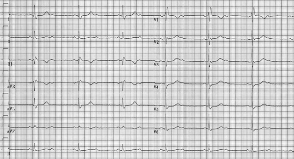
An 83-old-female patient presents to your outpatient clinic with a history of syncope. It has occurred three times in the last few months and there were no specific triggers. She is otherwise feeling well, has no other complaints and does not have any relevant medical history. Physical examination reveals no abnormalities except for a relatively slow heart rate (45 beats/min). Her ECG is presented in figure 1.

What is your diagnosis and what would you consider the most likely cause of her complaints?
Answer
The interpretation of the ECG is rather straightforward. The ventricular rhythm is slow (≤40 beats/ min) and at first glance one may consider this to result from a slow sinus rhythm. In fact, the sinus rhythm is 80 beats/min and 2:1 atrioventricular conduction is present. In the extremity leads the second P wave may be mistaken for a U wave, but in particular in lead V1 the second, blocked P wave is clearly visible. Conduction through the ventricles occurs with a right bundle branch block. Repolarisation is normal. Hence, this ECG is characterised by significant conduction abnormalities, i.e. second-degree AV block and right bundle branch block. Therefore, the most likely reason for her syncopes is a bradycardia and pacemaker therapy seems warranted.