File:E309.jpg

Original file (3,292 × 1,887 pixels, file size: 5.96 MB, MIME type: image/jpeg)
Summary
| Description |
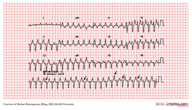
A 45 year old lady with palpitations and history of chronic renal failure Ventricular tachycardia A wide QRS tachycardia is VT until proven otherwise (1). Features suggesting VT include:- evidence of AV dissociation independent P waves (shown by arrows here) capture or fusion beats beat to beat variability of the QRS morphology very wide complexes (> 140 ms) the same morphology in tachycardia as in ventricular ectopics history of ischaemic heart disease absence of any rS, RS or Rs complexes in the chest leads (2) concordance (chest leads all positive or negative)
with permision from the site of Dr. Dean Jenkins |
|---|---|
| Category | |
| Source |
EKG World Encyclopedia http://cme.med.mcgill.ca/php/index.php , courtesy of Michael Rosengarten BEng, MD.McGill |
| Date |
2012 |
| Author |
Michael Rosengarten BEng, MD.McGill |
| Permission |
Creative Commons Attribution Noncommercial Share-Alike License |
File history
Click on a date/time to view the file as it appeared at that time.
| Date/Time | Thumbnail | Dimensions | User | Comment | |
|---|---|---|---|---|---|
| current | 09:38, 21 February 2012 |  | 3,292 × 1,887 (5.96 MB) | DarrelC (talk | contribs) |
You cannot overwrite this file.
File usage
The following page uses this file: