Answer MI 23
| This page is part of Cases and Examples
Previous ECG: MI 22 | Next ECG: This was the last MI case, return to Cases and Examples |
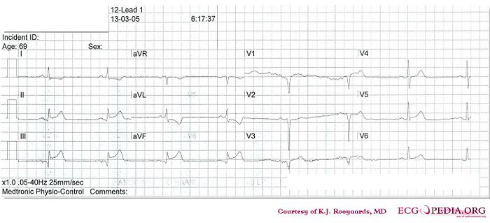
Where is this myocardial infarction located?

Answer
V3 is positioned at V4R.
- Following the 7+2 steps:
- Rhythm
- The ECG shows a regular rhythm. Every P wave is followed by a QRS complex. P waves are positive in I and AVF. Normal sinus rhythm
- Heart rate
- 40 bpm
- Conduction (PQ,QRS,QT)
- PQ: 160ms QRS: 90ms QT: 420ms QTc: 360ms
- Heartaxis
- QRS positive in I and AVF: normal heart axis
- P wave morphology
- Normal P wave morphology
- QRS morphology
- Pathologic Q waves in III and AVF. QS in V1 and V2. No ST elevation in V3 (=V4R).
- ST morphology
- ST elevation in II, III, AVF and V5. ST depression in I, AVL.
- Compare with the old ECG (not available, so skip this step)
- Conclusion?
- Rhythm
sinusbradycardia with inferior-lateral myocardial infarction.