Answer MI 21
| This page is part of Cases and Examples |
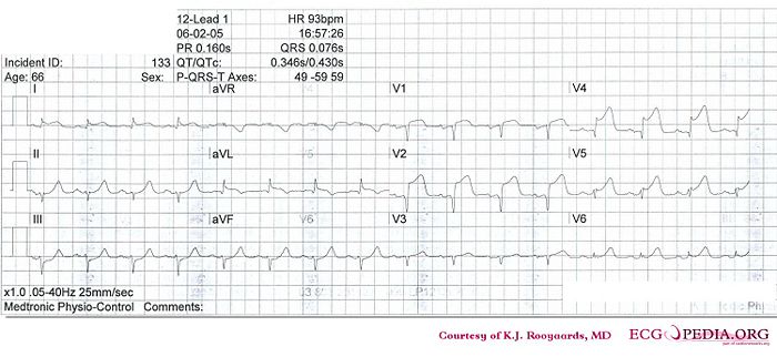
Where is this myocardial infarction located?

Answer
- Following the 7+2 steps:
- Rhythm
- The ECG shows a regular rhythm with normal P waves (positive in I, III and AVF, negative in AVR), followed by QRS complexes. Sinusrhythm
- Heart rate
- 90 bpm
- Conduction (PQ,QRS,QT)
- PQ: 160ms QRS: 80 ms QT: 340ms QTc: 416ms
- Heartaxis
- QRS positive in I and negative in II and AVF: left heart axis deviation
- P wave morphology
- P wave positive in II, III and AVF, biphasic in V1. The P waves have normal morphology.
- QRS morphology
- Narrow QRS. QS in V1-V2. Q in V4. Reduced R wave progression.
- ST morphology
- ST elevation in I, AVL, V1-V5 (V3=V4R and not elevated). ST depression in III and AVF.
- Compare with the old ECG (not available, so skip this step)
- Conclusion?
- Rhythm
Sinusrhythm with anteroseptal infarction. Ischemic vector is pointing upwards (ST depression in AVF), a sign of proximal LAD occlusion.