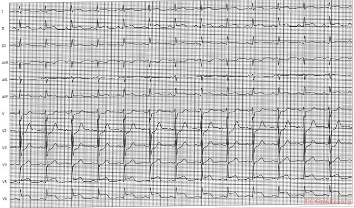
Answer MI 10
| This page is part of Cases and Examples |
Where is this myocardial infarction located?

Answer
Culprit lesion: RCX
- sinus rhythm
- about 77/min
- normal conduction
- intermediate axis
- normal p wave morphology
- No pathologic Q or LVH. Tall R in V2, V3.
- ST depression in V1, V4, tall R in V2. ST elevation in V5, V6. Some ST elevation in II, III and AVF.
- Conclusion: Inferior-lateral MI caused by an RCX occlusion.