Puzzle 2007 12 426 Answer
| Author(s) | A.A.M. Wilde | |
| NHJ edition: | 2007:06,231 | |
| These Rhythm Puzzles have been published in the Netherlands Heart Journal and are reproduced here under the prevailing creative commons license with permission from the publisher, Bohn Stafleu Van Loghum. | ||
| The ECG can be enlarged twice by clicking on the image and it's first enlargement | ||
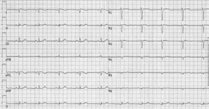
A 47-year-old lady presents with an ‘abnormal ECG’. In her family three daughters suffer from a long-QT syndrome type 2 and the responsible mutation has been identified in her husband and his family. An ECG was taken as part of a routine checkup (figure 1).
Is it abnormal and if so, what would your diagnosis be?
Answer
The ECG reveals a regular supraventricular rhythm of 60 beats/min. The P-wave morphology is abnormal (negative in leads 1 and aVL and positive in the inferior leads, suggesting a left superior atrial origin). Conduction intervals are normal. The electrical axis has a rightward shift and there are QS complexes in leads I and aVL. The QRS complexes in the left lateral leads have an abnormally low voltage (and slightly resemble a rightward placed ECG as obtained, for example, in the setting of an inferior myocardial infarction). The repolarisation is normal (QTc 414 ms, with a normal morphology). The combination of a negative P wave in leads 1 and aVL should raise two additional thoughts:
- accidental left-right exchange of the extremity leads and
- abnormal position of the heart.
In combination with the low-voltage left lateral leads, the latter possibility is by far the most likely, i.e. a rightward positioning of the heart: dextrocardia. Figure 2 shows a second ECG in which the precordial leads are placed at the right side of the thorax and that normalises the precordial leads. An exchange of the left and right arm electrodes would have normalised the whole ECG. Figure 3 shows the X-ray of the patient that confirms the dextrocardia. The family history of the long-QT interval in her offspring was already explained (segregation through the father’s line) and, based on her ECG, there is no reason to believe that she also suffers from a long-QT syndrome.
