Atrial Rhythm

Revision as of 15:18, 23 July 2007 by Drj (talk | contribs)
This is part of: Supraventricular Rhythms
Atrial Rhythm
{{{locatieafbeelding}}}
Atrial rate 50-100 bpm
Ventricular rate 1:1
Regularity regular
Origin atrial
P-wave present, but different from sinus rhythm
Effect of adenosine slows down
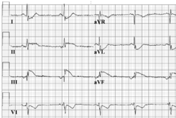
Example ECG: Atrial rhythm. In this example the sinus node stopped pacing due to ischemia during an atrial infarction. The p-wave is positive in I, negative in III and AVF. The atrial pacemaker is thus situated at the bottom of the right atrium, close to the AV node.
Example ECG2: {{{example2}}}

Atrial rhythm resembles sinusrhythm, but origins from a different atrial focus. It can be recognised by the abnormal configuration of the p-wave. Often the p-wave is negative in AVF, as is seen in the example.