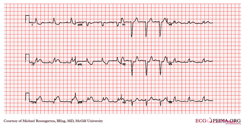
File:E319.jpg

Original file (3,004 × 1,599 pixels, file size: 4.47 MB, MIME type: image/jpeg)
Summary
| Description |
A 79 year old man with 5 hours of chest pain Acute myocardial infarction in the presence of left bundle branch block Features suggesting acute MI ST changes in the same direction as the QRS (as shown here) ST elevation more than you'd expect from LBBB alone (e.g. > 5 mm in leads V1 - 3 {not this case}) Q waves in two consecutive lateral leads (indicating anteroseptal MI) (ref. Sgarbossa EB et al, N Engl J Med 1996;334:481-7)
|
|---|---|
| Category | |
| Source |
EKG World Encyclopedia http://cme.med.mcgill.ca/php/index.php , courtesy of Michael Rosengarten BEng, MD.McGill |
| Date |
2012 |
| Author |
Michael Rosengarten BEng, MD.McGill |
| Permission |
Creative Commons Attribution Noncommercial Share-Alike License |
File history
Click on a date/time to view the file as it appeared at that time.
| Date/Time | Thumbnail | Dimensions | User | Comment | |
|---|---|---|---|---|---|
| current | 09:59, 21 February 2012 |  | 3,004 × 1,599 (4.47 MB) | DarrelC (talk | contribs) |
You cannot overwrite this file.
File usage
The following page uses this file: