File:E195.jpg

Size of this preview: 800 × 426 pixels. Other resolution: 3,004 × 1,599 pixels.
Original file (3,004 × 1,599 pixels, file size: 4.46 MB, MIME type: image/jpeg)
Summary
| Description |
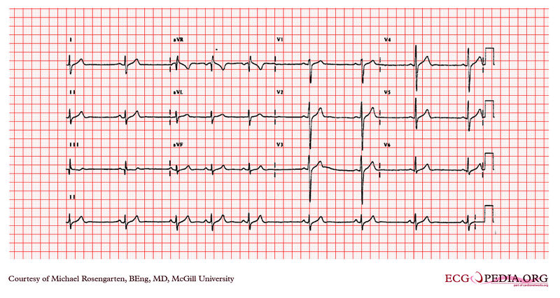
This EKG shows sinus rhythm, with a some what wide QRS and a right axis deviation. The S1 Q3 pattern suggest an isolated left posterior fasicular block. One should also consider right ventricular hypertrophy but there is no sign of right atrial abnormality, and his echocardiogram was normal. |
|---|---|
| Category | |
| Source |
EKG World Encyclopedia http://cme.med.mcgill.ca/php/index.php , courtesy of Michael Rosengarten BEng, MD.McGill |
| Date |
2012 |
| Author |
Michael Rosengarten BEng, MD.McGill |
| Permission |
Creative Commons Attribution Noncommercial Share-Alike License |
File history
Click on a date/time to view the file as it appeared at that time.
| Date/Time | Thumbnail | Dimensions | User | Comment | |
|---|---|---|---|---|---|
| current | 00:25, 21 February 2012 |  | 3,004 × 1,599 (4.46 MB) | DarrelC (talk | contribs) |
You cannot overwrite this file.
File usage
The following page uses this file: