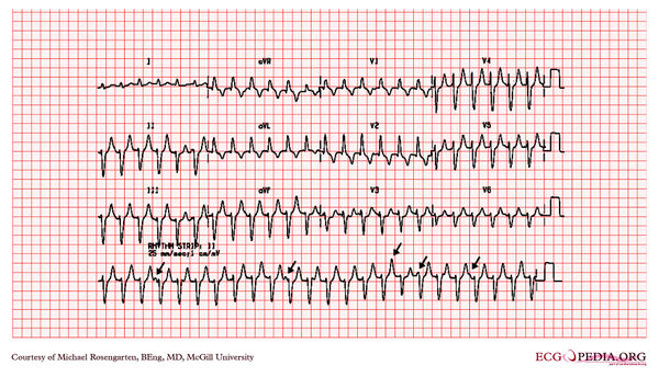
McGill Case 309
| This case report is kindly provided by Michael Rosengarten from McGill and is part of the McGill Cases. These cases come from the McGill EKG World Encyclopedia.
|

A 45 year old lady with palpitations and history of chronic renal failure Ventricular tachycardia A wide QRS tachycardia is VT until proven otherwise (1). Features suggesting VT include:- evidence of AV dissociation independent P waves (shown by arrows here) capture or fusion beats beat to beat variability of the QRS morphology very wide complexes (> 140 ms) the same morphology in tachycardia as in ventricular ectopics history of ischaemic heart disease absence of any rS, RS or Rs complexes in the chest leads (2) concordance (chest leads all positive or negative) 1) Griffith MJ, Garrat CJ, Mounsey P, Camm AJ. Ventricular tachycardia as the default diagnosis in broad complex tachycardia. Lancet. 1994;343:386- 2) Brugada P, Brugada J, Mont L, et al. A new approach to the differential diagnosis of a regular tachycardia with a wide QRS complex. Circulation. 1991;83:1649-1659 with permision from the site of Dr. Dean Jenkins
