...ker one month before. The pacer was programed to VVI mode and AAI mode and ECG recordings were made. Also a PA and lateral chest x-ray was taken. The ques
547 bytes (86 words) - 22:48, 19 February 2012
...logical examinations (X-ray, echo) were without abnormalities. Part of the ECG is shown in figure 1. Only the extremity leads are shown (standard calibrat
ms (rate slightly lower than 100 beats/min). The wide intervals on the ECG result from a blocked atrial impulse every fourth beat. The block is at the
2 KB (271 words) - 19:04, 3 August 2011
except for a relatively slow heart rate (45 beats/min). Her ECG is presented in figure 1.
644 bytes (97 words) - 19:55, 25 January 2010
 | File:E0003163.jpg |Description = This is an ECG strip and an audio recording from a patient checking his Medtronic VVI pace
(3,004 × 512 (1.06 MB)) - 00:41, 19 February 2012 |
[[Image:KJcasus6.jpg|700px|thumb|left|ECG MI 17. Click on image for enlargement.]]
**Compare with the old ECG (not available, so skip this step)
2 KB (287 words) - 20:22, 29 March 2012
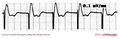 | File:E0003180.jpg ...ker one month before. The pacer was programed to VVI mode and AAI mode and ECG recordings were made. Also a PA and lateral chest x-ray was taken. The ques
(3,004 × 985 (730 KB)) - 01:30, 19 February 2012 |
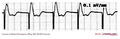 | File:E301.jpg ...ker one month before. The pacer was programed to VVI mode and AAI mode and ECG recordings were made. Also a PA and lateral chest x-ray was taken. The ques
(3,004 × 968 (718 KB)) - 08:16, 21 February 2012 |
...echocardiography, 24-h ambulatory Holter monitoring, and exercise testing. ECG Features of cardiac diseases detectable at pre-participation screening in y
...eased with age and level of exercise. In young amateur athletes they found ECG abnormalities in about 7%, a number that rose to 40% in "adult elite athlet
9 KB (1,320 words) - 09:21, 12 December 2011
heart failure. The ECG on admission is shown in
713 bytes (104 words) - 14:08, 19 May 2010
[[Image:Brugada.png|thumb|Typical ECG abnormalities in Brugada syndrome: ST elevation in V1-V3, without ischemia.
[[Image:Brugada_ecg_characteristics.svg|thumb| Typical ECG abnormalities in Brugada syndrome]]
8 KB (1,210 words) - 05:54, 22 May 2013
'''Q: ECG of a 28 year old man with atypical chestpain. (Lead V3 is at V4R position).
752 bytes (110 words) - 01:56, 17 May 2011
[[Image:SSS_ecg_001.jpg|thumb|ECG with Sick Sinus Syndrome. Rapid atrial fibrillation abruptly stops.]]
886 bytes (112 words) - 10:51, 25 April 2010
'''Question: What rhythm is shown on the ECG and what may be the cause?'''
644 bytes (105 words) - 09:15, 10 June 2012
...abnormalities, nor does laboratory investigation or echocardiography. Her ECG is presented: leads II, III, aVF, and V4 to V6
830 bytes (120 words) - 14:15, 19 May 2010
...logical examinations (X-ray, echo) were without abnormalities. Part of the ECG is shown in figure 1. Only the extremity leads are shown (standard calibrat
773 bytes (111 words) - 19:55, 25 January 2010
==ECG algorithms to differentiate wide QRS-complex tachycardias==
Several ECG algorithms have been developed to differentiate wide QRS-complex tachycardi
5 KB (750 words) - 19:01, 24 February 2013
...this knowledge it is quite simple to recognize normal sinus rhythm on the ECG.
...s it is of great importance. Arrhythmias include the most life-threatening ECG abnormalities. In most settings, however, the rhythm will be sinus.
2 KB (340 words) - 21:10, 14 January 2021
Image:E000549.jpg| Case 2b: ECG from the same patient before the MI occured.
817 bytes (138 words) - 18:00, 15 August 2011
ECG-characteristics:<cite>Wung</cite>
...sion or even elevation in II, III, and AVF) the distal LAD is occluded. An ECG that does not show any ST depression sugggests an occlusion after the origi
3 KB (506 words) - 10:00, 8 October 2014
[[Image:ami0006.jpg|700px|thumb|left|ECG MI 6]]
855 bytes (131 words) - 18:35, 2 April 2011