File:E314.jpg

Original file (3,292 × 1,887 pixels, file size: 5.91 MB, MIME type: image/jpeg)
Summary
| Description |
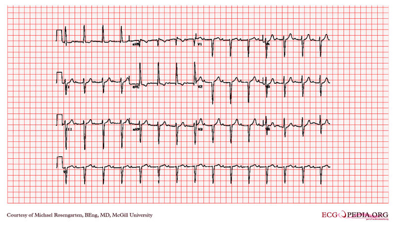
An 84 year old lady with hypertension There are a number of abnormalities here. left anterior hemiblock QRS axis more left than -30 degrees initial R wave in the inferior leads (II, III and aVF) absence of any other cause of left axis deviation left ventricular hypertrophy In the presence of left anterior hemiblock the diagnostic criteria of LVH are changed. Rosenbaum suggested that an S wave in lead III deeper than 15 mm as predictive of LVH. long PR interval (also called first degree heart block) PR interval longer than 0.2 seconds left atrial hypertrophy M shaped P wave in lead II P wave duration > 0.11 seconds terminal negative component to the P wave in lead V1 with permision from the site of Dr. Dean Jenkins |
|---|---|
| Category | |
| Source |
EKG World Encyclopedia http://cme.med.mcgill.ca/php/index.php , courtesy of Michael Rosengarten BEng, MD.McGill |
| Date |
2012 |
| Author |
Michael Rosengarten BEng, MD.McGill |
| Permission |
Creative Commons Attribution Noncommercial Share-Alike License |
File history
Click on a date/time to view the file as it appeared at that time.
| Date/Time | Thumbnail | Dimensions | User | Comment | |
|---|---|---|---|---|---|
| current | 09:56, 21 February 2012 |  | 3,292 × 1,887 (5.91 MB) | DarrelC (talk | contribs) |
You cannot overwrite this file.
File usage
The following page uses this file: