McGill Case 53: Difference between revisions
No edit summary |
No edit summary |
| (One intermediate revision by the same user not shown) | |
(No difference)
| |
Latest revision as of 08:34, 13 February 2012
| This case report is kindly provided by Michael Rosengarten from McGill and is part of the McGill Cases. These cases come from the McGill EKG World Encyclopedia.
|

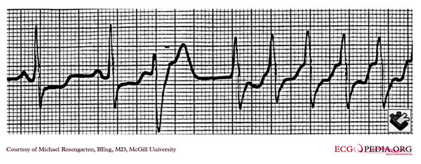
This is a rhythm strip recorded by telemetry on a 70 year old patent. The major time lines are 0.2 of a second. Note that the tachycardia is initiated with a premature ventricular beat with the retrograde P'wave sitting on the up sloping section of the T wave. There is a long P'R interval to the next QRS with is the beginning of the tachycardia at a rate of about 160/min. With this tachycardia the patient had chest pain and a blood pressure of 70mm systolic. A coronary angiogram found severe triple vessel coronary disease.

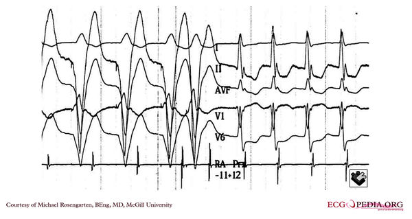
This is the same patient in the electrophysiology laboratory. A ventricular catheter is used to stimulate the ventricle and a right atrial (RA) catheter is used to record the atrial activity. The patient has been premedicated with digoxin 0.25mg/day. The tracing shows four surface leads and one intra cardiac recording. Supraventricular tachycardia is induced with a train of ventricular beats and a final premature beat. Note that the last stimulated beat has a shorter pacing interval and results in a longer interval between it and the next RA deflection. This then sets off AVNRT with the RA activity now closely associated with the QRS. The short-long interval between the initiating beat and the first QRS of the tachycardia is similar to that of the spontaneous tracing. The long interval suggest conduction down a slow anterograde pathway which is typical of AVNRT.

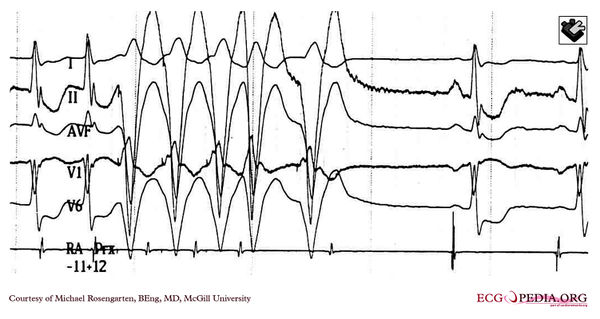
This is a tracing with four surface leads and one intracavitary right atrial (RA) recording. The tracing began when the patient was in AVNRT. The rate of the tachycardia was 135/min . This rate is slower that the patient's initial rate. This may be due to the effect of digitalis. The digitalis could also explain the large ST segment changes, or these could be due to the tachycardia itself or ischemia. Note the ventricular pacing train is interrupted by a premature spontaneous or reentrant ventricular complex which fails to conduct in the retrograde direction and results in termination of the arrhythmia.
