Wide Complexes Intervening Regular Sinus Rhythm: Difference between revisions

From ECGpedia
Jump to navigation Jump to search
m (New page: {{NHJ| |mainauthor= '''A.A.M. Wilde''' |edition= 2006:12,436 }} Figure 1|thumb A 28-year-old, wheelchair bound, female patient with Friedreich’s at...)
 
mNo edit summary
Line 18: Line 18:


[[Puzzle_2006_12_436_Answer|Answer]]
[[Puzzle_2006_12_436_Answer|Answer]]
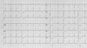
The ECG shows sinus rhythm at a rate of 90 beats/
min. Conduction intervals are normal (PR 120 ms,
QRS 90 ms). Aspecific repolarisation abnormalities
(terminal negative T waves in the left lateral and inferior
leads) are observed. These are more frequently seen in
patients with Friedreich’s ataxia and do not seem to have
any particular meaning.
There are four wide QRS complexes, one recorded
in the extremity leads and three in the precordial leads.
Given the similar configuration in lead II (rhythm strip
at the bottom) the 1st aberrant QRS complex is
probably similar to the 3rd aberrant QRS complex.
These complexes are preceded by a short PR interval
and ventricular pre-excitation is suggested. The 2nd and
4th aberrant complexes are wider and not preceded
by a discernable P wave. Both are followed by a complete
compensatory pause suggesting that these complexes
have a ventricular origin. The question that
subsequently arises is whether all four wide QRS
complexes are ventricular extrasystoles or whether both
pre-excited complexes and ventricular premature beats
are present.
If pre-excitation is suggested then the approximate
site of pre-excitation should be given. In this case the
suggested Δ-wave is positive in leads I, II and III but
negative in leads aVR and aVL. The latter combination
is actually impossible with pre-excitation and this makes
a ventricular extrasystole originating from the right
ventricular outflow track, fused with ventricular
activation through the normal conduction system
(following the regular P-wave) more likely. Indeed,
the initial part of the other wide complexes is similar
to the initial morphology of the ‘pre-excited’ complexes.
A more likely diagnosis therefore is ventricular
extrasystoles with different coupling intervals (and
fusion in the 1st and 3rd complex).

Revision as of 19:25, 8 October 2007

Author(s) A.A.M. Wilde
NHJ edition: 2006:12,436
These Rhythm Puzzles have been published in the Netherlands Heart Journal and are reproduced here under the prevailing creative commons license with permission from the publisher, Bohn Stafleu Van Loghum.
The ECG can be enlarged twice by clicking on the image and it's first enlargement
Figure 1

A 28-year-old, wheelchair bound, female patient with Friedreich’s ataxia presents with an irregular heartbeat. She has no history of syncope or dizziness and no other complaints. Physical examination of the heart reveals no abnormalities. There are neither signs of left nor right heart failure. Her pulse is in principle regular, but occasionally some irregularities are observed. The ECG is depicted in figure 1. An echo is normal.

What is your diagnosis?
Answer