File:Brugada syndrome type1 example5.png
Jump to navigation
Jump to search
Size of this preview: 800 × 473 pixels. Other resolution: 3,300 × 1,950 pixels.
Original file (3,300 × 1,950 pixels, file size: 364 KB, MIME type: image/png)
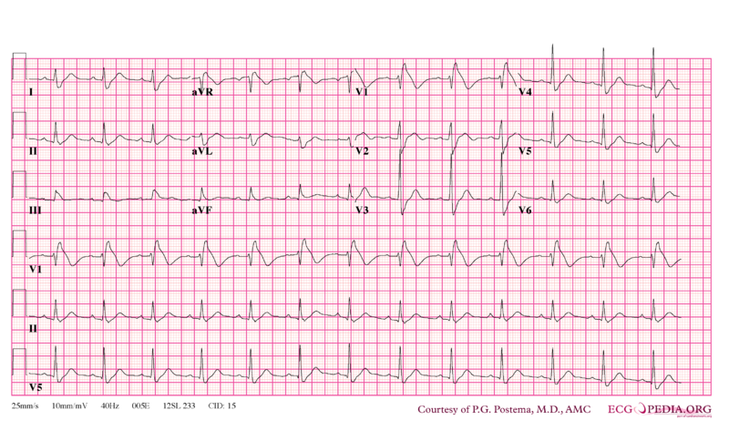
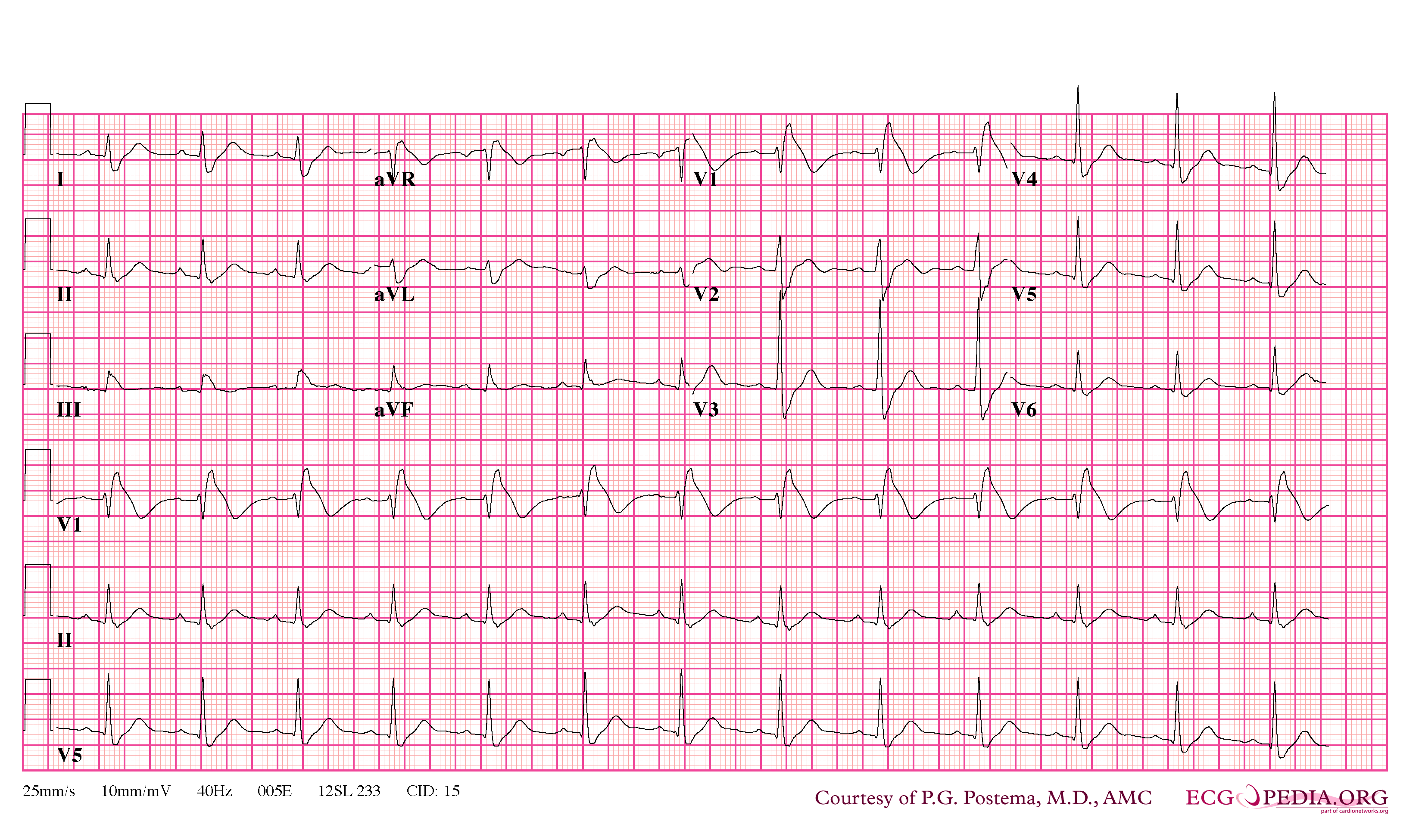
There is only (!) a Type I morphology in V1, so officially this would not meet the criteria for a Brugada Syndrome ECG. Leads should have placed one intercostal space above V1 and V2 to reach the diagnosis (which was later done). Notwithstanding that the Type I morphology in V1 is overwhelming and is accompanied by frequently seen wide S waves in the lateral and inferior leads.
File history
Click on a date/time to view the file as it appeared at that time.
| Date/Time | Thumbnail | Dimensions | User | Comment | |
|---|---|---|---|---|---|
| current | 10:04, 10 April 2010 | | 3,300 × 1,950 (364 KB) | (username removed) |
File usage
The following page uses this file: