Example 26
Jump to navigation
Jump to search
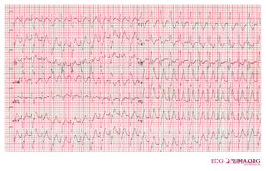
This patient presented with a broad complex tachycardia, shown in figure 1.
The figure shows a broad complex tachycardia 160/min with RBBB configuration and extreme heart axis. Following the broad complex tachycardia flowchart:
- Fusion beats: no
- Absence of precordial RS: no
- R-to-S > 100ms: no
- AV-dissociation: yes
Heart axis and AV-dissociation make a ventricular tachycardia most likely.
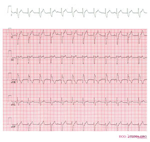
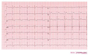
- 7.5 mg verapamil was administered, which slowed down the VT and ultimately the patient converted to sinusrhythm


